TCM: Tissue Composition Module
Investigational Use Only
Get the best tools for understanding bone status, structure and change
The Mindways Tissue Composition Module (TCM) extends our QCT Pro™ software to produce estimates of visceral adipose tissue (VAT), subcutaneous adipose tissue (SAT), intramuscular adipose tissue (IMAT) and lean tissue mass. Increased VAT has been associated with increased risk of cardiovascular disease and type 2 diabetes, while fat infiltration of muscle tissue and loss of lean muscle tissue mass (sarcopenia) is related to impaired muscle strength which is predictive of incident disability and all-cause mortality in the elderly.
Volumetric Assessment
In comparison to area-based assessments of tissue composition, such as those provided by DXA, CT's ability to resolve the spatial distribution of various tissue types in three dimensions provides a more detailed view of the distribution of fat and fat infiltration of other tissue types.
Calibration
The same calibration technology used by Mindways for the estimation of bone mineral density can be used to quantify fat content and muscle density. In particular, the TCM is compatible with CT images acquired either with or without a CT calibration phantom imaged with a patient and thus is suitable for use in both prospective and retrospective studies. The use of calibrated tissue measurements facilitates standardization of measurements across CT scanners and CT scan technique as well as quantification of the amount of fat in other tissues such as average fat in muscle (IMAT).
Semi-Automated
TCM incorporates a semi-automatic analysis mode that significantly expedites the tissue analysis relative to manual segmenation of CT images used in many tissue quantification studies.
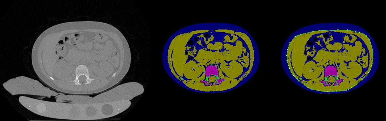
Abdominal CT
Abdominal CT images can be processed with TCM to estimate cross-sectional areas and volumes of lean and fat tissue types, including visceral adipose tissue area (VAT).
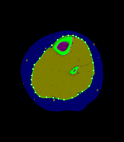
Peripheral CT